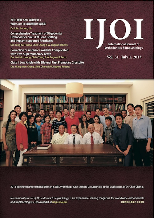
IJOI Vol. 31

Comprehensive Treatment of Oligodontia: Orthodontics, Sinus Lift Bone Grafting, and Implant-supported Prostheses
Huang TK, Chang CH, Roberts WE
This report describes the interdisciplinary treatment of an acquired malocclusion in an adult male that was associated with oligodontia and anterior crossbite. Six premolars were congenitally missing, four in the maxilla and two in the mandible, resulting in multiple, intermaxillary edentulous areas. In the mandibular arch, all spaces were closed and incisors were retracted to correct the anterior crossbite. In the maxillary arch, space was consolidated to develop implant sites to replace the the missing first premolars. Due to inadequate bone height bilaterally, the edentulous areas were restored with dental implants placed with simultaneous sinus lift, bone grafting procedures. Prosthodontic restoration was then completed using implant-supported crowns. Occlusal function, dental esthetics and the smile-line were markedly improved. (Int J Orthod Implantol 2013;31:16-39.)
Key words: oligodontia, self ligation bracket, sinus lift, bone grafting, lateral approach, osteotome technique, sinus membrane perforation, implant-supported prosthesis
Download Article
Correction of Anterior Crossbite Complicated with Two Supernumerary Teeth
Huang YH, Chang CH, Roberts WE
History and Etiology
A 12-year-1-month-old girl was accompanied by her parents for evaluation of a crowded dentition (Fig. 1). The chief complaint was an unesthetic smile due to a maxillary anterior crossbite and crowding. There was no other contributory medical or dental history. Clinical examination revealed an anterior crossbite, with blocked out right and left maxillary canines, and a bilateral class I molar relationship (Figs. 2 and 3). The patient was treated to an acceptable result as documented in Figs. (4-6); however, the treatment of the anterior crossbite (Fig. 7) was complicated by the presence of two supernumerary teeth (mesiodens) in the premaxillary region (Figs. 8). Figures 9 and 10 provide a direct comparison of the cephalometric and panoramic radiographs before and after treatment, respectively. Figure 11 shows the superimposition of the tracings for the before and after treatment cephalometric radiographs. (Int J Orthod Implantol 2013;31:44-60)
Class II Low Angle with Bilateral First Premolars Crossbite
Chang HW, Chang CH, Roberts WE
History and Etiology
A young female, aged 27-years-old (Fig. 1), presented with a chief complaint of her irregular teeth arrangement and protruding upper anterior teeth (Figs. 2-3). There was no contributory medical or dental history. The clinical exam indicated that the bilateral first premolars were crossbite and a large overbite was noticed (Fig. 2). Her pre-treatment facial profile showed a straight profile with an acceptable soft tissue E-line projection. The pre-treatment intraoral photographs and study models revealed a bilateral end-on Class II molar relationship. The lower dental midline was shifted to the right side. No contributing habits were evident. The patient was treated to an acceptable result as documented in (Figs. 4-9). The cephalometric and panoramic radiographs document the pre-treatment conditions (Fig. 7) and the post-treatment results (Fig. 8). Superimposed cephalometric tracings document the treatment achieved (Fig. 9). The details for diagnosis and treatment will be discussed below. (Int J Orthod Implantol 2013;31:62-75)
Compromised Treatment for an Asymmetric Class II/III Mutilated Malocclusion with Facial Asymmetry
Chang HW, Chang CH, Roberts WE
History and Etiology
A 23-year-6-month-old male presented for orthodontic consultation with chief complaints of irregular dentition, mandible shift and facial asymmetry (Fig. 1). There was no contributory medical or dental history. A clinical exam revealed that the permanent (Int J Orthod Implantol 2013;32:64-77) [… read more]
阻生齒軟組織處理面面觀
Soft Tissue Considerations
for The Management of Impactions
Su B, Hsu YL, Chang CH, Roberts WE
治療阻生齒與其它一般的矯正病患治療時間相比,往往需要較長的治療時間,這類病患的治療也涉及牙周手術與矯正科的參與,從術前診斷,正確的治療計劃,阻生齒暴露術式的選擇與矯正的互相配合, 有時甚至還需要矯正中以及矯正後的簡單手術與, 才能進一步達到美學的要求。
我們在臨床上遇到阻生齒的機會其實不低,平均約為 1% ,其中上下顎比例約為四比一;上顎阻生齒中發生在顎側的比例較高;術式考量也與頰側完全不同。以下為各位醫師介紹我們在臨床上常用的四種術式。 [… read more]