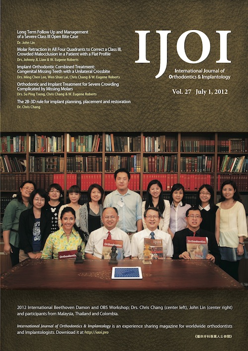
IJOI Vol. 27

Long Term Follow Up and Management of a Severe Class III Open Bite Case
Lin JJ
Introduction
The patient presented with seemingly simple Class III asymmetry with a labially block out left upper canine. The initial treatment plan indicated traditional edgewise orthodontic appliances for better alignment. The patient would then stay in long term follow up until the active growth period was completed and be ready for second stage correction of the asymmetric malocclusion. (Int J Orthod Implantol 2012;27:4-16)
Molar Retraction in All Four Quadrants to Correct a Class III, Crowded Malocclusion in a Patient with a Flat Profile
Liaw JL, Roberts WE
HISTORY AND ETIOLOGY
A 26 year old male patient presented for consultation with a chief complaint of dental protrusion. He asked for extraction treatment to reduce the perceived protrusion. However clinical examination revealed a relatively retrusive maxilla and straight profile, with no sign of dental protrusion. Apparently the maxillary incisor prominence, due to severe crowding, led to his mistaken impression of “protrusion” (Figs. 1-3). The preliminary diagnosis was a mild skeletal Class III relationship, with dental compensation, that resulted in flaring of the upper incisors and lingual tipping of the lower incisors. Based on the examination and history, the etiology of the malocclusion appeared to be primarily genetic. (Int J Orthod Implantol 2012;27:20-33)
Orthodontic and Implant Treatment for Severe Crowding Complicated by Missing Molars
Tseng SP, Chang CH, Roberts WE
History and Etiology
A 33-year-old female was referred by her dentist for orthodontic consultation to evaluate her Class II Division 2, mutilated dentition (Fig. 1). Bilateral miniscrews were evident in the infrazygomatic crest areas, that had been placed by her dentist, prior to the decision to send the patient for specialty evaluation. The patient’s chief concern was an irregular dentition, with two missing teeth in the lower left posterior area (Figs. 1-2). No other contributing medical or dental history was reported. (Int J Orthod Implantol 2012;27:34-51)
Atypical Extraction of Adult Orthodontic Treatment
Chang MJ, Chang CH, Roberts WE
History and Etiology
A 27-years-old female was referred by her dentist for orthodontic consultation (Fig. 1). Her chief concern was maxillary anterior crowding and missing mandibular teeth (Figures 2, 3). There were no contributory medical problems. Clinical exam indicated that the bilateral maxillary lateral incisors were in cross-bite and mandibular left 1st molar and right 1st premolar were missing (Fig. 2). The patient was treated to an acceptable result as documented in Figs. 4-9. The cephalometric and panoramic radiographs document the pre-treatment conditions (Fig. 7) and the post-treatment results (Fig. 8). The cephalometric tracings before and after treatment are superimposed in Fig. 9. The details for diagnosis and treatment will be discussed below. (Int J Orthod Implantol 2012;27:52-63)
Implant-Orthodontic Combined Treatment: Congenital Missing Teeth with a Unilateral Crossbite
Lee MC, Lai WS, Chang CH, Roberts WE
HISTORY AND ETIOLOGY
A 23-year-11-month-old male was referred by his dentist for orthodontic consultation (Fig. 1). His chief concern was dental spacing and multiple teeth in crossbite (Figs. 2-3). There was no other contributory medical or dental history. Clinical exam indicated multiple missing teeth in the maxilla: both lateral incisors, right 2nd premolar, and right 1st molar. The lower right 2nd premolar was also missing (Fig. 2). A treatment plan combining orthodontics, prosthetic implants and implant-supported prostheses was proposed to correct the skeletal and dental problems. (Int J Orthod Implantol 2012;27:66-81)
Congenital Missing of Mandibular Incisors with Class I Malocclusion
Hung J, Chang CH, Roberts WE
HISTORY AND ETIOLOGY
A 21 year old female was evaluated for maxillary dental crowding (Figs. 1-3). She had received orthodontic treatment for 1 year at the age of 12, but was dissatisfied with the long-term result. The initial clinical exam revealed a Class I molar relationship bilaterally, associated wth maxillary anterior crowding and two missing mandibular incisors. The overjet was 6mm, and overbite was 4mm. The maxillary dental midline was shifted 1mm to the right of the facial and mandibular midlines. Oral soft tissues, frena and gingival health were all within normal limits. There was no history of dental trauma or aberrant oral habits. There was no other contributory medical or dental history. The patient desired comprehensive orthodontic treatment to achieve an ideal alignment of the entire dentition, which was achieved as documented in Figs. 4-6. (Int J Orthod Implantol 2012;27:82-93)
The 2B-3D rule for implant planning, placement and restoration
Chang CH
What is biologic width?
Is there a golden rule for implant planning, placement and restoration as the Newton’s laws of motion for force prediction? In order to answer this question, one needs to refer back to the biologic system which the implant site attempts to mimic. (Int J Orthod Implantol 2012;27:96-101)
Rehabilitation of the Maxillary Arch with Implant-supported Fixed Restorations Guided by the Most Apical Buccal Bone Level in the Esthetic Zone: A Clinical Report (Chinese translation)
Su B, Rojas-Vizcaya F